Fig. 2.
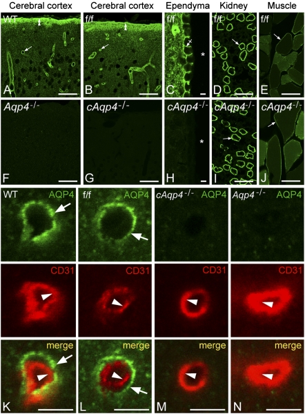
Immunofluorescence micrographs showing distribution of AQP4. The AQP4 labeling (green) pattern in cerebrum of WT (A) and f/f mice (B and C) was indistinguishable. Note distinct immunosignal around vessels (arrows), underneath the pia (double arrowheads), and along the basolateral membrane of ependymocytes (crossed arrow). Asterisks, third ventricle. In cerebrum of constitutive (F) and cAqp4−/− (G and H) mice, AQP4 immunoreactivity was absent. Conditional deletion of Aqp4, however, did not affect the labeling pattern in muscle and kidney (compare I and J with D and F; arrows indicate kidney collecting duct and sarcolemma, respectively). Double labeling of obliquely cut capillaries with the endothelial marker CD31 (red) revealed that the AQP4 labeling (green) associated with microvessels in WT and f/f mice was peripheral to the endothelium (arrowhead), corresponding to perivascular astrocytic endfeet (arrows; K and L). No detectable signal was seen over endothelial cells in either genotype (K and N). (Scale bars: A, B, D, E, F, G, I, and J, 50 μm; C, H, and K–N, 5 μm.)

