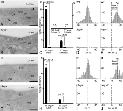
Fig. 3.
Subcellular distribution of AQP4 immunogold reactivity. (A) Electron micrograph from WT mouse showing AQP4 immunogold particles along the astrocyte (Ast) endfoot membrane. End, endothelial cell; Peri, pericyte. Arrowheads indicate astrocyte endfoot membrane (1) and the endothelial membranes facing astrocyte (2), pericyte (3), and lumen (4). (B) AQP4 labeling was absent in constitutive Aqp4 knockout mice (Aqp4−/− mice). (C) Quantitative analysis of the density of gold particles over the membrane domains indicated in A. The same symbols (1–4) are used for easy reference. Gold particles within 23.5 nm from a given membrane were allocated to that membrane. The AQP4 signal over the Ast endfoot membrane was significantly higher in WT than in Aqp4−/− mice. The two genotypes did not differ in AQP4 densities over endothelial membranes facing pericytes (End peri) and lumen (End lu), indicating absence of AQP4 in these membrane domains. The AQP4 signal over endothelial membranes next to astrocyte endfeet (End ast) was higher in WT than in Aqp4−/− mice. (D and E, Upper) Analysis in WT animals of the distribution of gold particles perpendicular to the astrocyte endfoot membrane (Ast, D) or to the endothelial membrane facing astrocytes (End ast, E) revealed peak immunogold signal coinciding with former membrane. When analysis was performed with abluminal endothelial membrane as reference (E) the peak was skewed corresponding to the thickness of the basal lamina (indicated with bar; Results). (D and E, Lower) No peaks were seen in Aqp4−/− mice. (F) AQP4 immunogold labeling pattern in controls (f/f) was similar to that of WT mice (A). (G) AQP4 immunoreactivity was absent in glial-conditional Aqp4 knockout mice (cAqp4−/− mice). (H) The linear densities of AQP4 signaling gold particles over astrocytic and apposed endothelial membranes showed a pronounced reduction in cAqp4−/− vs. litter controls (f/f). (I and J) Same design as in D and E, respectively, but f/f controls in lieu of WT and cAqp4−/− mice in lieu of Aqp4−/− mice. Perpendicular distribution of gold particles showed absence of peaks after glial-conditional deletion of Aqp4, confirming that the immunosignals seen in f/f mice reside in glia. (Scale bars: A, B, F, and G, 200 nm.)

