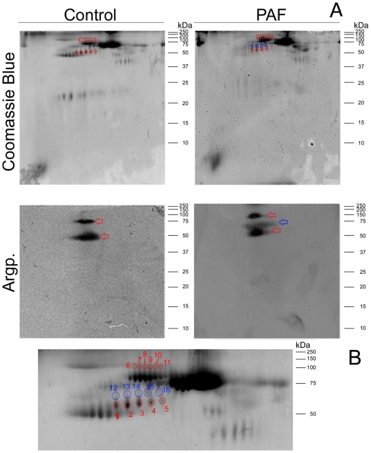
Figure 2. Two dimensional electrophoresis of control and FAP plasma proteins stained with Coomasie and Western blot analysis of argpyrmidine modified proteins.
(A) Full gel image; (B) Zoom image of the gel area at which protein spots were removed. Assigned protein spots were excised for protein identifications (Table 1). Red marks correspond to common glycated proteins and blue marks correspond to protein glycated differentially in FAP individuals.

