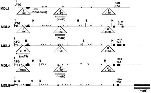
Figure 3.
Comparison of the major features of the MDL1 to MDL5 cDNAs, including their initiation codons (ATG), termination codons (TAA/TGA), signal sequences (open rectangles), putative FAD-binding sites (closed ovals), insertions (closed rectangles), and potential N-glycosylation sites (v). Triangles indicate where the ORFs of the mdl1 to mdl4 genes are interrupted in the genome by three introns. The intron sizes are shown in parentheses within each triangle. The positions of fragments used as probes in Southern-blot analysis are indicated by gray bars. B, E, and H denote BamHI, EcoRI, and HincII restriction sites, respectively. Not indicated are additional EcoRI (−594) and HincII (−1941) sites in the mdl3 promoter region.

