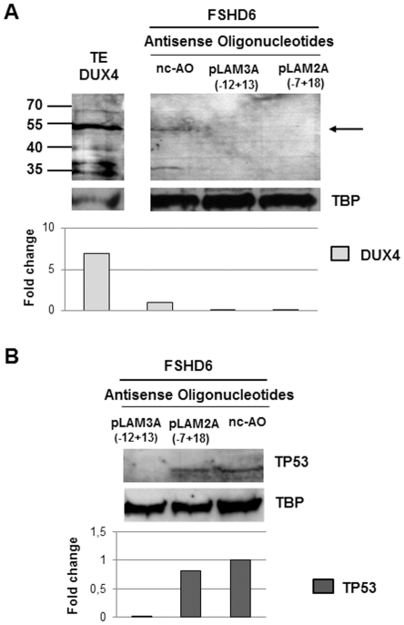
Figure 6. Efficiency of AOs pLAM2A (−7+18) and pLAM3A (−12+13) in suppressing endogenous DUX4 and TP53 expression in primary FSHD myotubes.
(A) 105 primary FSHD myoblasts were seeded in 35 mm culture dishes. The next day, cells were transfected with either the negative control AO mGMCSF3A(−5+20) (nc-AO, 600 nM) or AOs pLAM2A (−7+18) (50 nM) or pLAM3A (−12+13) (150 nM). Differentiation was induced 4 hours after transfection and cells were harvested 72 hours later. Nuclear extracts were prepared and 20 µg of proteins were separated by electrophoresis (12% PAGE-SDS), transferred to a Western blot and DUX4 was immunodetected with 9A12 MAb. The antibodies were then stripped, and the same membrane used for immunodetection of TBP. The methodology for the Western blot is shown in the legend to Fig. 5 . TE-DUX4: positive control, 5 µg protein extract of TE671 cells transfected with a pCIneo-DUX4 expression vector (B) Immunodetection of TP53 with specific primary antibody and appropriate secondary antibody as described in the legend to Fig. 3 on Western blot prepared with protein extracts of cells used in the above experiment (6A). A densitometry of the immunoreactive bands was performed. Data are normalized to TBP levels in each sample.

