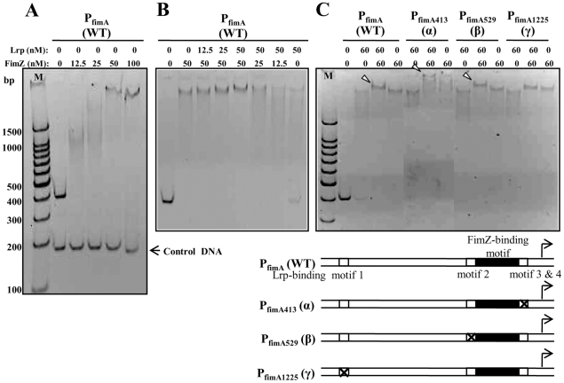
Figure 7. Binding of the purified Lrp and FimZ proteins to PfimA.
(A) Binding of FimZ to PfimA(WT). (B) Binding of Lrp and FimZ to PfimA(WT). (C) Binding of Lrp and FimZ to PfimA(WT), PfimA413 (α), PfimA529 (β), or PfimA1225 (γ). The white arrowheads indicate super-shifted Lrp-PfimA-FimZ complexes. To enhance resolution of the super-shifted DNA-protein complexes, the running time of the polyacrylamide gel in panel C was extended from 1 h (used in panels A and B) to 2 h. Schematic diagrams of the wild-type and mutant fimA promoters are shown under the gel.

