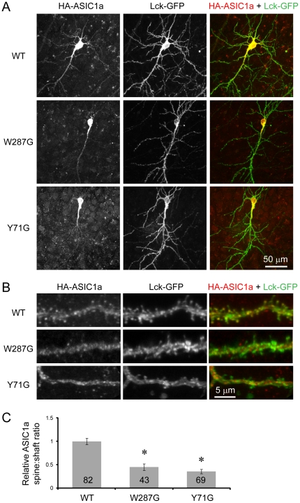
Figure 1. Mutating Tyr71 and Trp287 reduces dendritic targeting of ASIC1a.
ASIC2-/- hippocampal slices were co-transfected with HA-tagged wild-type ASIC1a, W287G or Y71G mutants together with a membrane targeted Lck-GFP [22]. Localization of transfected ASIC1a was visualized by immunofluorescence using an anti-HA antibody. (A) Confocal images showing the overall distribution of ASIC1a in transfected neurons. Left panel shows ASIC1a localization, middle panel shows GFP fluorescence, right panel shows the merged image (HA immunofluorescence in red and GFP fluorescence in green). Note the presence of wild-type ASIC1a in most dendritic branches, while mutant ASIC1a localized primarily in the cell body and proximal dendrites. (B) High magnification view of a segment of apical dendrite. Note the presence of wild-type but not mutant ASIC1a in most dendritic spines. (C) Quantification of relative ASIC1a spine:shaft ratio. The ratio of ASIC1a spine:shaft was normalized to that of the membrane-targeted Lck-GFP. The normalized ratio of wild-type ASIC1a was set arbitrarily to 1. Numbers on the bar indicate the total number of spines quantified from 5 wild-type, 4 71G, and 5 287G transfected neurons from two separate experiments. Asterisks indicate significant differences (P<0.0001, unbalanced ANOVA).

