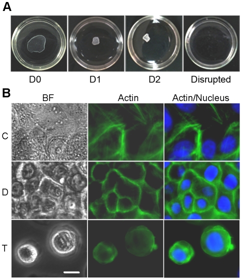
Figure 1. Loss of cell-substrate and cell-cell contact results in reorganization of actin cytoskeleton.
(A) Images of dispase-lifted cell sheets at zero, one and two days post-dispase lifting, and disrupted by the mechanical shear test, in 35 mm dishes. (B) Immortalized human keratinocytes were divided cells into three treatment groups: control (C), confluent cells that maintained in a cell culture dish, had both cell-cell and cell-substrate contact; dispase-lifted (D), confluent cells that lifted as an intact cell sheet, maintained cell-cell contact but lost cell-substrate contact, and trypsinized (T), cells that trypsinized and suspended as single cells, had neither cell-cell nor cell-substrate contact. Representative images of bright field (left panels), actin stain (middle panels), and actin merged with nucleus stain (right panels) in untreated control cells (top panels), dispase-lifted cell sheet (middle panels), and trypsinized cells (bottom panels) show attenuation or loss of non-junctional stress fibers in the dispase-lifted cell sheet and loss of actin organization in the trypsinized cells. Further, cells in the dispase-lifted cell sheet shrunk in the horizontal plane upon release from the substrate. Green: actin, Blue: nucleus. Scale bar: 10 um.

