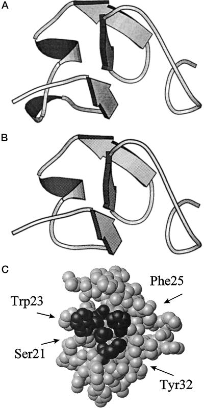
Figure 7.
Three-dimensional models of hevein (A) and SN-HLPf (B). Strands of the β-sheet and stretches of α-helix are represented by arrows and ribbons, respectively. N-terminal ends of polypeptide chains are on the right. Cartoons were rendered using the program Molscript (Kraulis, 1991). C, Space-filling model of SN-HLPf showing the exposed Ser-21, Trp-23, Phe-25, and Tyr-32 residues (dark gray), which build the putative binding site of the protein. This model was generated using the MOLMOL program (Koradi et al., 1996).

