
Fig. 9.
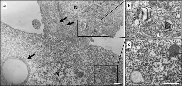
Terfenadine induces ultrastructural features of autophagy. A375 cells were treated with 10 μM TEF for 8 h and processed for electron microscopy. a The induction of autophagy is manifested by massive vacuolization of the cytoplasm. Arrows indicate autophagic vacuoles different in size. b Note the double membrane or the multimembrane structure of the autophagic vacuoles. c In the same cell, immature autophagic vacuoles characterized by an electron density equivalent to the cytoplasm coexist with late vesicles, in which catabolic processes have been already started (characterized by an increased electron density). Bars 1 μm