
Figure 8.
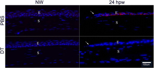
Phospho-AKT staining in the healing corneal epithelium of DT-depleted mouse corneas. Cryostat sections of PBS- or DT-injected mouse corneas, with wounding (24 hpw) or without wounding (NW), were immunostained with antibody against phospho-AKT (Ser473). Photographs show merged images of immunofluorescence of phospho-AKT (red) and nuclear staining of DAPI (blue). Arrows mark the leading edge of migration corneal epithelial cells. The figures are representatives of four corneas per condition from two independent experiments. E, epithelium; S, stroma.