


Figure 2.
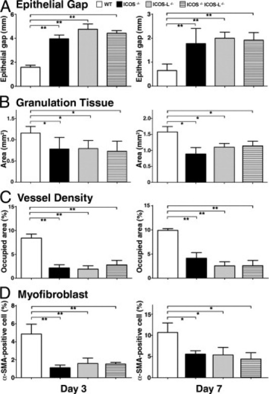
Keratinocyte migration, granulation tissue formation, angiogenesis, and myofibroblast proliferation in wild-type (WT) and mutant mice. A: The distance between the migrating edges of keratinocytes under the eschar (epithelial gap) and (B) the area of granulation tissue, were both measured in tissue sections. C: Vessel density was determined as CD31-positive areas, and (D) the frequency of myofibroblasts was identified as the percentage of α-SMA–positive cells among fibroblastic cells. Each histogram shows the mean (+SEM) results obtained from at least 10 mice of each group. *P < 0.05, **P < 0.01.