
Figure 1.
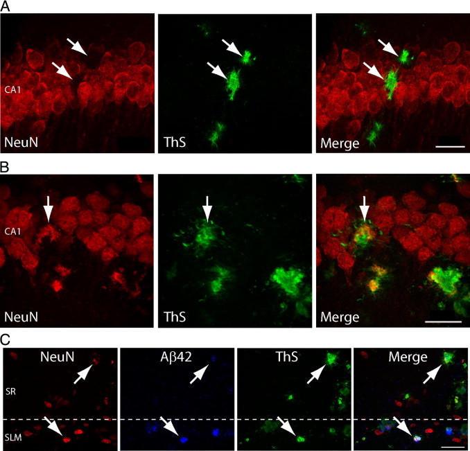
Thioflavin S (ThS) positive plaques colocalize with neuronal marker NeuN in CA1 hippocampus. A: The 5-month-old Tg19959 mouse reveals ThS (green) positive β-amyloid (Aβ) plaques within the CA1 pyramidal cell body layer. At this age, areas that are labeled with ThS (arrows) are near but not directly colabeled with neuronal marker NeuN (red; arrows). B: In contrast, ThS and NeuN colocalize (merged arrows) in the CA1 pyramidal cell body layer in 3-month-old Tg19959 mice. C: Colocalization (arrows) of ThS, Aβ42 (blue), and NeuN is also seen in synaptic layers of the hippocampus, stratum radiatum, and stratum lacunosum moleculare in 3-month-old Tg19959 mice, supporting that GABAergic interneurons are also a site of ThS positive Aβ fibrils. Scale bars: 25 μm (A, B), 50 μm (C). Animations of the high-resolution three-dimensional reconstructions (B, C) are shown in Supplemental Videos S1 and S2 (available at http://ajp.amjpathol.org), respectively.