
Figure 2.
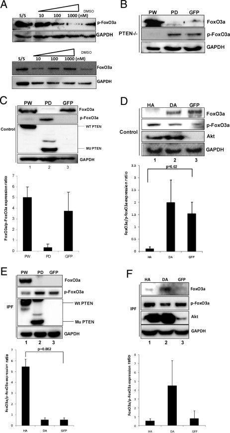
Inhibition of Akt decreases p-FoxO3a levels in IPF fibroblasts. Serum-starved IPF fibroblasts were pre-incubated with an Akt inhibitor, ranging from 10 to 1000 nmol/L, and plated on collagen matrix for 60 minutes. A: p-FoxO3a (top) and FoxO3a (bottom) levels were examined by Western blot analysis. Glyceraldehyde-3-phosphate dehydrogenase (GAPDH) was used as a loading control. Dimethylsulfoxide (DMSO)–treated cells were used as a control. B: PTEN−/− cells were infected with adenoviral constructs expressing wild-type PTEN (PW), phosphatase-deleted PTEN (PD), or empty vector (GFP), and cells were cultured on polymerized collagen for 24 hours. Western blot analysis was performed to measure FoxO3a, p-FoxO3a, and GAPDH protein levels. C: Top: Control fibroblasts infected with PW, PD, or empty vector GFP were cultured on polymerized collagen for 24 hours, and FoxO3a, p-FoxO3, and PTEN levels were measured. GAPDH was used as a loading control. Bottom: The FoxO3a/p-FoxO3a expression ratio in control fibroblasts infected with PW, PD, or empty vector (GFP) was measured by densitometry. D: Top: Control human lung fibroblasts infected with an adenovirus expressing HA, DA, or empty vector (GFP) were cultured on polymerized collagen for 24 hours, and Western blot analysis was performed to measure FoxO3a, p-FoxO3a, Akt, and GAPDH levels. Bottom: The FoxO3a/p-FoxO3a expression ratio was quantified by densitometry. E: Top: IPF fibroblasts overexpressing PW, PD, or empty vector (GFP) were cultured on polymerized collagen for 24 hours, and FoxO3a, p-FoxO3a, and PTEN levels were measured. GAPDH was used as a loading control. Bottom: The FoxO3a/p-FoxO3a expression ratio was quantified by densitometry. F: Top: IPF fibroblasts infected with an adenovirus expressing HA, DA, or empty vector (GFP) were cultured on polymerized collagen for 24 hours, and Western blot analysis was performed to measure FoxO3a, p-FoxO3a, Akt, and GAPDH levels. Bottom: The FoxO3a/p-FoxO3a protein expression ratio was measured by densitometry.