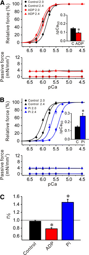
Fig. 2.

Effects of MgADP or Pi on Ca2+ sensitivity and t 1/2 in rabbit psoas muscle fibers; SL 2.0 and 2.4 μm. a Effect of 3 mM MgADP on force–pCa curves (top) and passive force (bottom). Black solid lines −MgADP, red solid lines +MgADP. Inset comparison of ΔpCa50 in the absence and presence of MgADP. C control without MgADP. *P < 0.05; n = 6. b Effect of 20 mM Pi on force–pCa curves (top) and passive force (bottom). Black solid lines −Pi (same as in a), blue solid lines +Pi. Inset comparison of ΔpCa50 in the absence and presence of Pi. *P < 0.05; n = 6. Passive force was not significantly altered by MgADP or Pi at either 2.0 or 2.4 μm (a, b). c Comparison of t 1/2 in the absence and the presence of 3 mM MgADP or 20 mM Pi. *P < 0.05 compared with control; n = 6
