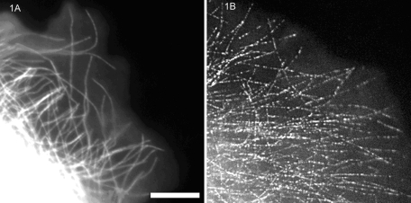
FIGURE 1:
Comparison of diffraction-limited fluorescent images recorded with a cooled CCD camera and 1.4–numerical aperture objective of microtubules in the lamella of a migrating newt lung epithelial cell injected with X-rhodamine–labeled tubulin. (A) Ten percent labeled tubulin and (B) 0.25% labeled tubulin in the cytosol. Scale bar, 10 μm. (Reproduced with permission from Waterman-Storer CM, Salmon ED (1999). Fluorescent speckle microscopy of microtubules: how low can you go? FASEB J 13(Suppl 2), S225–S230.)

