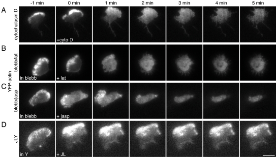
FIGURE 2:
JLY preserves steady-state actin organization in HL-60 cells. HL-60 cells stably expressing YFP-tagged actin were plated on fibronectin-coated slides and imaged in DIC and oblique fluorescence illumination. Cells were pretreated with 100 nM fMLP and (B and C) 50 μM blebbistatin or (D) 10 μM Y27632 for 10 min before imaging. At t = 0 min, (A) 10 μM cytochalasin D, (B) 500 nM latrunculin B, (C) 1 μM jasplakinolide, or (B) 8 μM jasplakinolide and 5 μM latrunculin B were added. Images are representative of a minimum of five cells. Scale bar: 10 μm.

