FIGURE 1:
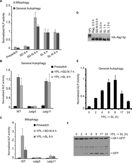
Induction of autophagy upon switch from a rich medium to a minimal, non-starvation medium. Cells were grown in rich medium (YPL) to log phase before switch to nitrogen-starvation medium (SD-N or SL-N). Non-starvation media (SD and SL) were used as controls. At the indicated time points, general autophagy or mitophagy was measured by ALP activity assay or by Western blot using Idh1-GFP as the reporter for mitophagy. IDH1 encodes a mitochondrial matrix protein. (A) As expected, autophagy was induced in starvation media (SD-N and SL-N) but not in the control medium (SD). Surprisingly, autophagy was also significantly induced upon switch to SL medium. (B) NNS-induced general autophagy and nitrogen-starvation-induced general autophagy were inhibited in Δatg5 cells, and not affected in Δatg11 cells. (C) NNS-induced mitophagy and nitrogen-starvation-induced mitophagy were inhibited in both Δatg5 and Δatg11 cells. (D) Dephosphorylation of Atg13p triggered by autophagy-inducing conditions. Rapamycin treatment and nitrogen starvation led to dephosphorylation of Atg13p as reported previously (Kamada et al., 2000). Atg13p was also dephosphorylated upon switch to SL medium. Cells were grown in YPL to log phase before switch to rapamycin-containing (0.2 μg/ml) YPL medium, nitrogen-starvation medium (SD-N), or SL medium. At the indicated time points, 5 OD cells were quenched by mixing with TCA to a final concentration of 5-6% and incubated on ice for at least 5 min before centrifugation. Cell pellets were then subject to whole-cell TCA extraction and analyzed by immunoblotting. Rap., rapamycin. (E and F) Time course experiments revealed that autophagic activity became detectable after 2–4 h, peaked at approximately 6–8 h, and started to decrease after 17–24 h following switch to SL medium. In (A–C and E), data represent averages of three to five samples with error bars for standard deviations.

