Figure 3.

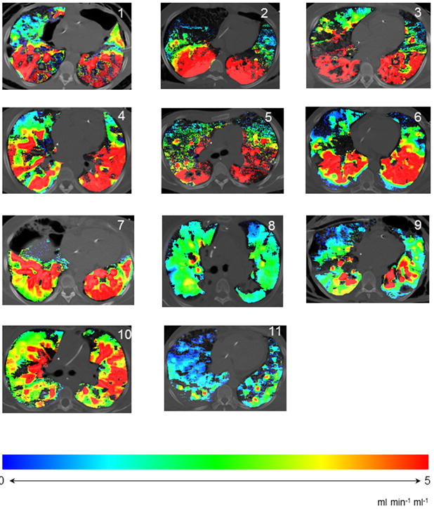
Color-maps, showing distribution of perfusion throughout and axial slice of lung in patients with ARDS. Spectrum changing from blue to red indicates increasing perfusion.

Color-maps, showing distribution of perfusion throughout and axial slice of lung in patients with ARDS. Spectrum changing from blue to red indicates increasing perfusion.