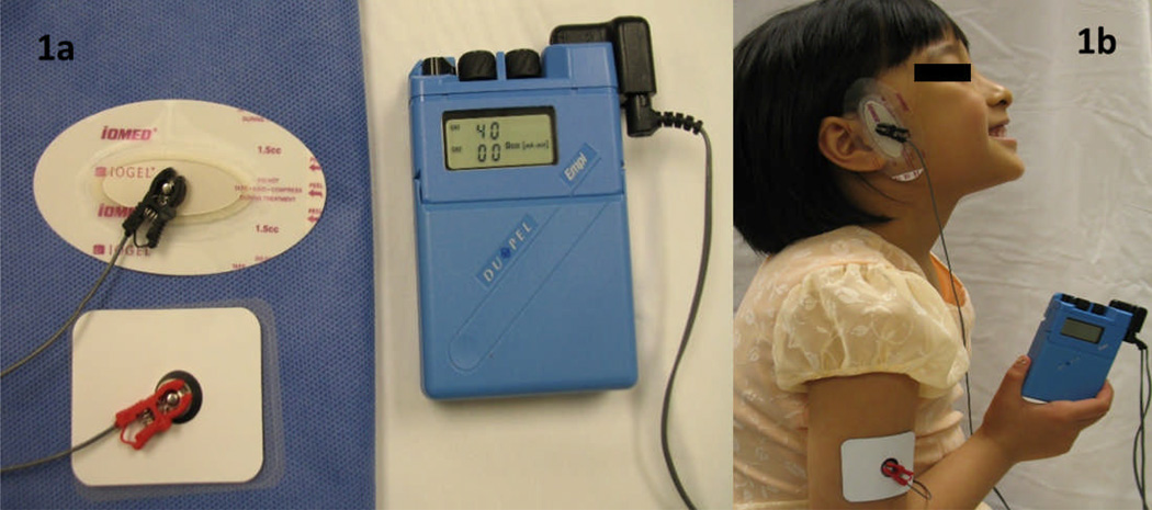
Figure 1. Dexamethasone iontophoresis of the temporomandibular joint.
Panel 1a depicts the iontophoresis equipment with its two bipolar electrodes. On the top is the oval delivery electrode which has the dexamethasone reservoir directly below the clamp. On the bottom is the square dispersive electrode. The iontophoresis device shows the total current dose to be administered. The dials on top of the iontophoresis device are used to adjust the level of current flow intensity.
Panel 1b shows the placement of the two electrodes during DIP sessions. The delivery electrode is placed on the involved TMJ and the dispersive electrode on the upper arm at the same side of the treated TMJ.

