
Figure 5. TARPs are expressed in OPCs.
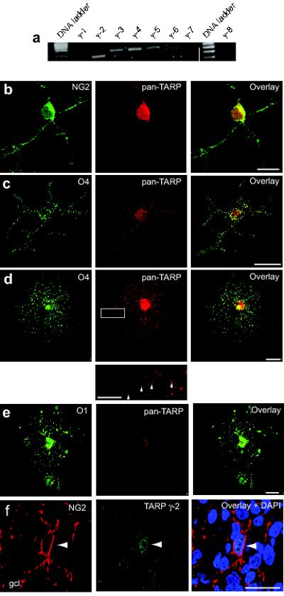
(a) RT-PCR analysis of TARP expression in the rat optic nerve. mRNA for γ-2, γ-3, γ-4, γ-5 and the TARP-related protein γ-6 was detected. (b-d) Representative confocal images showing labeling of (b) NG2+ cells, (c) O4+ cells, and (d) pre-myelinating OPCs, with anti-pan-TARP (red), anti-NG2 (green), anti-O4 (green) antibodies. Note the punctate TARP labeling (indicated by arrowheads) along the processes of the pre-myelinating OPC (inset, from white rectangle in d). (e) Representative confocal images of oligodendrocytes identified with anti-O1 (green). The cells exhibited reduced TARP immunoreactivity compared with that seen with pre-myelinating OPCs. Labeling similar to that shown in b-e was seen in 8-20 cells of each type across 12 separate cultures. Scale bars 25 μm (10 μm, inset). (f) Confocal images of a representative sagittal section of cerebellar cortex from a P7 rat, showing labeling of the granule cell layer (gcl) with anti-NG2 (red) and anti-pan-TARP (green) antibodies and nuclear staining with DAPI (blue). Scale bar 25μm. Arrowhead indicates presumptive NG2+ OPC.