Fig. 1.
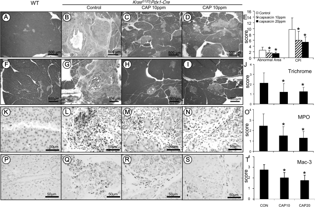
Analysis of chronic pancreatitis using the approaches of histopathology, immunohistochemistry and histochemistry. Histopathology in the pancreatic tissue of KrasG12D/Pdx1-Cre mice: (A) Pancreas in wild-type control mice showing morphologically normal pancreatic parenchyma (acini and islets as well interlobular ducts). (B) Extensive chronic pancreatitis in KrasG12D/Pdx1-Cre mice fed AIN-76A diet alone showing loss of acini and stromal fibrosis. (C and D) Minimal to mild chronic pancreatitis in mice fed 10 or 20 p.p.m. capsaicin-supplemented diet, respectively. (E) Semiquantitative analysis (histogram) of chronic pancreatitis based on the extent of acinar loss, stromal fibrosis and inflammatory cell infiltration. There was a statistically significant difference in mice fed AIN6A diet alone compared with those given capsaicin-supplemented diets (P < 0.05). Trichrome stain highlighting stromal fibrosis in the pancreas: (F) Trichrome stained fibroconnective tissue (blue color) only in the interlobular areas of a normal pancreas in wild-type control mice. (G) KrasG12D/Pdx1-Cre mice fed AIN-76A diet alone showing extensive fibrosis in pancreatic parenchyma. (H and I) mice fed 10 or 20 p.p.m. capsaicin-supplemented diet exhibiting much less stromal fibrosis in the pancreas. (J) Semiquantitative analysis (histogram) of the extent of trichrome stain-labeled stromal fibrosis revealing statistically significant differences in mice fed capsaicin-supplemented diets compared with those given AIN-76A diet alone (P < 0.05). MPO-labeled neutrophils in the pancreas: (K) Pancreas from wild-type control mice showing no MPO-labeled neutrophils. (L) KrasG12D/Pdx1-Cre mice fed AIN-76A diet alone displaying intense MPO-labeled neutrophils in the areas of pancreatitis. (M and N) Mice fed 10 or 20 p.p.m. capsaicin-supplemented diet exhibiting decreased MPO-positive neutrophils. (O) Semiquantitative analysis (histogram) of the MPO-labeled inflammatory cells showing a statistically significant difference in mice fed capsaicin-supplemented diets compared with those given AIN-76A diet alone (P < 0.05). Mac-3-labeled macrophages in the pancreas: (P) Pancreas from wild-type control mice showing no Mac-3-labeled inflammatory cells. (Q) KrasG12D/Pdx1-Cre mice fed AIN-76A diet alone displaying intense Mac-3-labeled macrophage in the areas of pancreatitis. (R andS) Mice fed 10 or 20 p.p.m. capsaicin-supplemented diet exhibiting decreased Mac-3-positive macrophages. (T) Semiquantitative analysis (histogram) of the Mac-3-labeled macrophages showing a statistically significant difference in mice fed capsaicin-supplemented diets compared with those given AIN-76A diet alone (P < 0.05).

