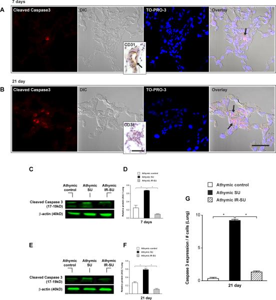
Figure 6.
IR of athymic rats leads to reduced pulmonary vascular endothelial cell apoptosis. (A) Immunofluorescent images of serial lung tissue sections for cleaved caspase 3 expression (arrow) at d7 after SU5416 administration. Differential interference contrast (DIC) represents small lung vessel histology and inset shows CD31 expression in small lung vessel (n=8/group). (B) Immunofluorescent images of serial lung tissue sections for cleaved caspase 3 expression (arrow) at d21 after SU5416 administration. DIC image shows an almost occluded vessel and inset shows absent CD31 staining inside the lung vessel wall (n=8/group). (C,D,E,F) Western blot analysis from whole lung lysate for cleaved caspase 3 protein levels at d7 and d21 (n=8/group). (G) Quantitation of activated caspase 3 positive endothelial cells in precapillary pulmonary arteries in lung tissue sections at d21 (n=4/group). Data are shown as means with error bars representing SEM. * p<0.05. Scale bar: (A,B)=50μm; insets=25μm.

