
Figure 6.
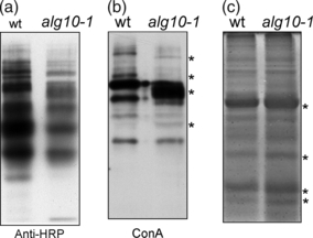
alg10-1 displays differences in N-glycosylation.
(a) Immunoblot analysis of total proteins extracted from wild-type (wt) and alg10-1 leaves. Proteins were subjected to SDS-PAGE under reducing conditions and blots were analyzed using anti-horseradish peroxidase (anti-HRP) antibodies, which recognize complex N-glycans with a β1,2-xylose and core α1,3-fucose residues.
(b) Proteins were subjected to SDS-PAGE under reducing conditions and blots were analyzed using the lectin concanavalin A (ConA).
(c) Coomassie brilliant blue staining of total protein extracts. Asterisks indicate bands that differ between wild-type and alg10-1.