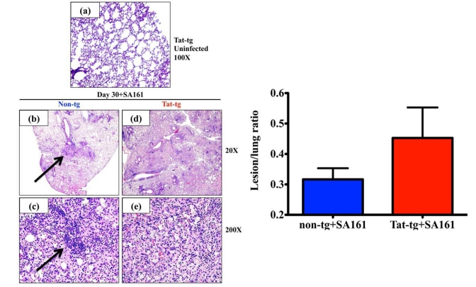
Fig. (2).
Increased lung pathology in Tat–tg mice infected with M. tuberculosis W-Beijing SA161 compared to non-tg control mice. (a) Representative photomicrograph of H&E-stained lungs from an uninfected Tat-tg mouse. Total magnification a=100x. (b-e) Lung sections from all 4 mice per group infected with a low dose aerosol of W-Beijing SA161 M. tuberculosis 30 days post infection were examined and a representative photomicrographs of H&E-stained lungs from a non-tg (b) and (c) or a Tat-tg mouse (d and e) are shown. Total magnification b, d=20x; c, e=200x. (f) H&E sections were analyzed; total lung area from all 4 mice were measured and compared to areas affected by lesions. The percent lung involvement is reported; SA161-infected Tat-tg mice (red bar), non-tg mice (blue bar).

