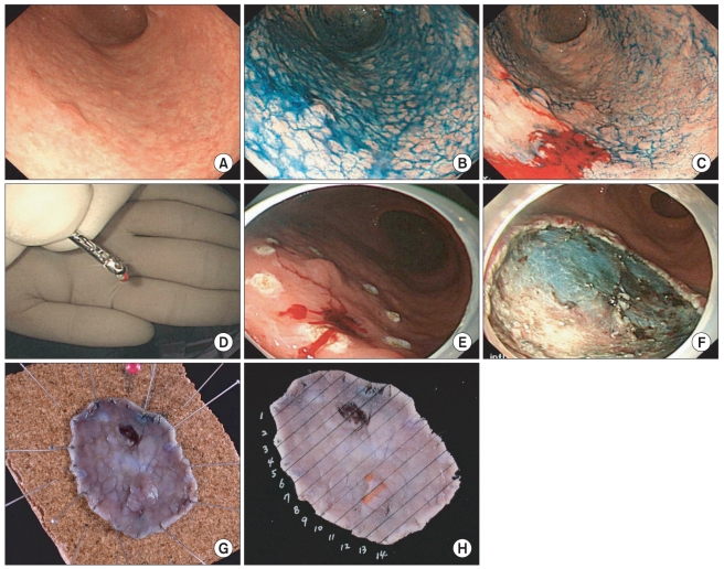
Fig. 2.
A representative case of gastric tubular adenoma in which frozen section biopsy was performed before endoscopic submucosal dissection. (A) A slightly elevated gastric lesion with unclear margin in white light was noticed in the lower third stomach. (B) The lateral spread of the lesion was still obscure despite of chromoendoscopy using acetic acid and indigo carmine. (C) A frozen section biopsy was performed 5mm distal to expected lateral extent of the lesion, which was selected by the experienced endoscopist (Cho JY), because the lesions showed disconcordance in the assessment of lateral spread between two endoscopists. (D) The outer surface of the specimen was marked with special ink (the Davidson Marking System® Bradely product Inc.) to provide a pathologist (Jin SY) orientation of specimens. (E) We marked several spots near the site, where frozen section biopsy was done, because frozen section revealed chronic gastritis with intestinal metaplasia. (F) En bloc resection of the lesion was performed. (G) A site of frozen section biopsy was observed in the resected specimen. (H) Pathologic mapping showed curatively resected tubular adenoma (The orange line indicated the lesion, tubular adenoma).

