Figure.
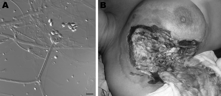
A) Sporangiophore (center) and sporangiospores of Apophysomyces variabilis fungi. Scale bar = 10 μm. B) Clinical manifestations in a woman infected with A. variabilis fungi in the upper part of the chest and the breast. A color version of this figure is available online (www.cdc.gov/EID/content/17/1/134-F.htm).

