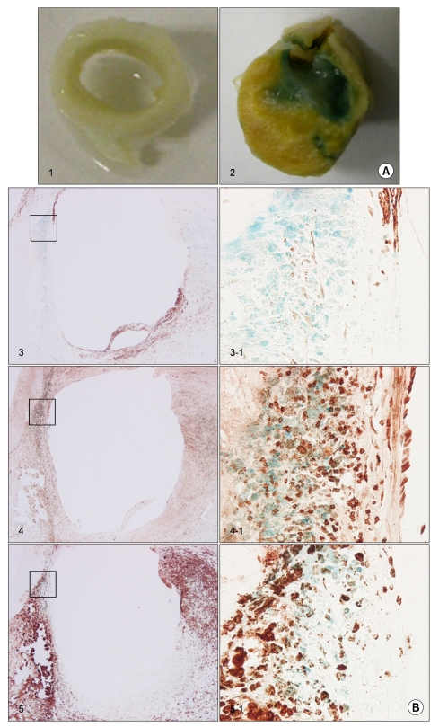
Fig. 2.
β-galactosidase (β-gal) activity in various arterial specimens (A) and β-gal and inmmunohistochemical staining in atherosclerotic plaques (B). (A) Photographs of the various arteries stained for β-gal activity (non-atherosclerotic iliac artery (1); endarterectomized carotid atheroma (2)). (B) The serial sections of the luminal layer of endarterectomized carotid atheroma stained for β-gal activity and immunohistochemistry stains for α-smooth muscle actin (3), CD3 (4), and CD68 (5). The (3), (4), and (5) were doble stained with β-gal and each antibody. The results were the same for experiment five (Magnification: ×40 for (3), (4), and (5); ×200 for (3-1), (4-1), and (5-1)).

