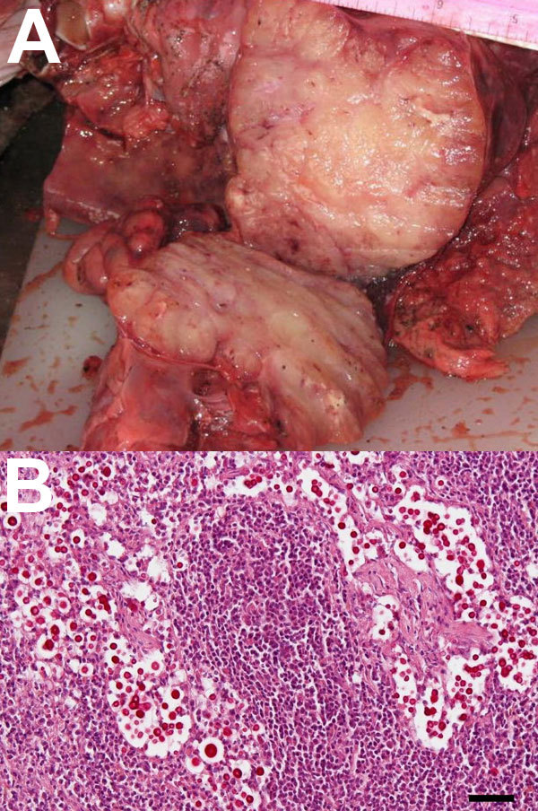
Figure A1.

A) Enlarged mediastinal lymph nodes of a stranded, pregnant, harbor porpoise (Phocoena phocoena) infected with Cryptococcus gattii that was transmitted to its fetus. B) Mucicarmine–stained sections of fetal mediastinal lymph node, showing C. gattii extracellular yeast aggregates (original magnification ×20). Scale bar = 50 μm.
