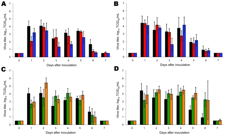
Figure 2.
Virus shedding from the nose and throat of ferrets inoculated with wild-type and reassortant pandemic (H1N1) 2009 viruses. Virus shedding from nose (A, C) and throat (B, D) is shown for pandemic (H1N1) 2009–seasonal influenza (H1N1) (A, B) and pandemic (H1N1) 2009–seasonal influenza (H3N2) (C, D) reassortant viruses. Black, wild-type pandemic (H1N1) 2009; red, pandemic (H1N1) 2009–seasonal influenza (H1N1) basic polymerase (PB) 2 acidic polymerase; blue, pandemic (H1N1) 2009–seasonal influenza (H1N1) PB2; green, pandemic (H1N1) 2009–seasonal influenza (H3N2) PB1 neuraminidase (NA); orange, pandemic (H1N1)–seasonal influenza (H3N2) NA. Geometric mean titers are shown; error bars indicate SD. The lower limit of detection is 0.5 log10 50% tissue culture infective dose/mL (TCID50/mL). After day 3, only 3 animals remained in each group.

