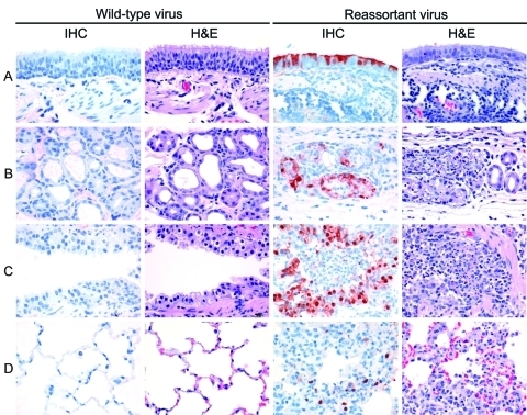
Figure 4.
Examples of virus antigen expression and severity of lesions in different tissues of the lungs of ferrets. A) Bronchial surface; B) bronchial submucosal gland; C) bronchiole; D) alveolus. Two of 3 ferrets inoculated with wild-type pandemic (H1N1) 2009 virus had neither virus antigen expression (first column) nor associated lesions (second column) in the lung at day 3 postinoculation. In contrast, all 3 ferrets inoculated with reassortant pandemic (H1N1) 2009–seasonal influenza (H3N2) virus neuraminidase had virus antigen expression in bronchi, bronchial submucosal glands, bronchioles, and alveoli (third column), associated with epithelial degeneration and necrosis and infiltration of inflammatory cells, predominantly neutrophils (fourth column). IHC, immunohistochemistry; H&E, hematoxylin and eosin stain. Original magnification ×400.

