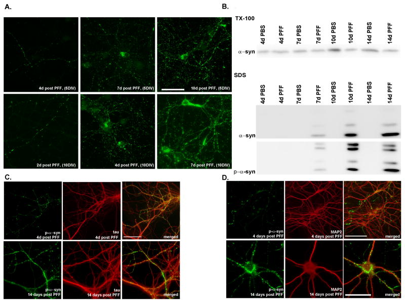
Figure 4. Time dependence of aggregate formation.
A. Top row: α-syn-hWT pffs were added to DIV5 neurons, and fixed either 4, 7 or 10 days later. Small puncta corresponding to neuritic p-α-syn were visible 4 days after pff addition, and by 7 days, neuritic p-α-syn levels increased, and accumulations were visible in some cell bodies. Ten days following addition of pffs, p-α-syn was seen throughout the neurites as small puncta, longer fibrous structures, and as somal accumulations. Bottom row: α-syn-hWT pffs were added to DIV10 neurons when α-syn expression at the presynaptic terminal is higher. Pathology progresses more quickly and are detectable at 2 days post-pff addition and aggregates in the cell bodies as early as 4 days post-pff addition. Scale bar = 50 μm. B. Immunoblots of DIV5 neurons sequentially extracted with 1% Tx-100 and 2% SDS, 4, 7, 10 and 14 days following PBS or α-syn-hWT pff addition. Over time, soluble α-syn was reduced with a concomitant increase in total and p-α-syn in the Tx-100-insoluble fraction. C, D. Double immunofluorescence for p-α-syn and the axonal marker, mouse tau (T49) (C). or the dendritic marker, MAP2 (D). P-α-syn predominantly colocalized with tau but not MAP2 4 days after pff addition. Two weeks after pff addition, aggregates were found in axons, cell bodies and dendrites where they colocalized with MAP2. Scale bar = 20 μm. See also Supplementary Figure 2.

