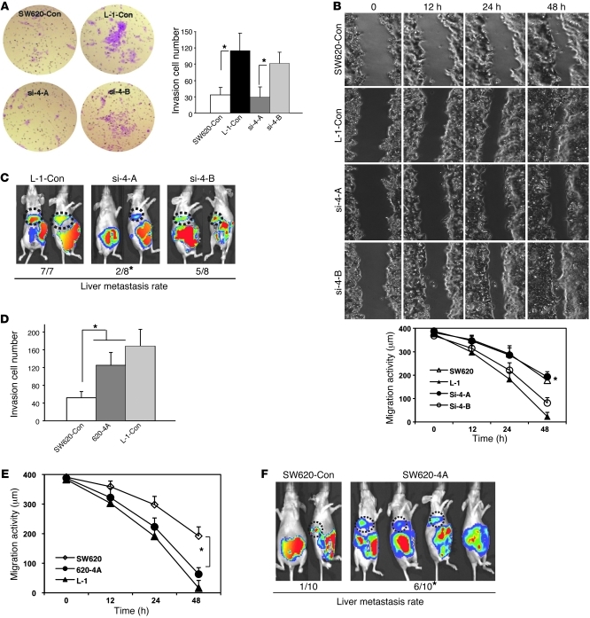
Figure 2. Four genes are essential in mediating colorectal cancer liver metastasis.
(A) Images show the invasion activity of the hepatic metastasis L-1 cell line with knocked down group A and group B genes using retroviral shRNAs (si-4A, si-4B, upper panel). Cells migrating to the lower side of the Transwell filter were counted per HPF in 3 fields (lower panel). Each assay was repeated at least twice. Original magnification, ×200. *P < 0.05. (B) Images show the migration activity of the hepatic metastases L-1 cell line with knocked down group A genes using wound healing assay (upper panel). The distance of cell migration was calculated at 3 locations (lower panel). Con, control. (C) Representative images of the IVIS luciferase signal in the mice inoculated with colorectal cancer cells preinfected with 2 groups of retroviral shRNAs. The hepatic metastasis rate is indicated at the bottom. *P < 0.05 compared with L-1–control cells. (D) Invasion activity of the hepatic metastasis L-1 cell line overexpressing the group A genes. (E) Migration activity of the hepatic metastases L-1 cell line overexpressing the group A genes. (F) Representative images of the luciferase signal in mice inoculated with colorectal cancer cells expressing the group A genes. The hepatic metastasis rate is indicated at the bottom. *P < 0.05 compared with SW620-control cells. Bars show the mean value of the representative results from 3 experiments, each conducted in duplicate (± SD).

