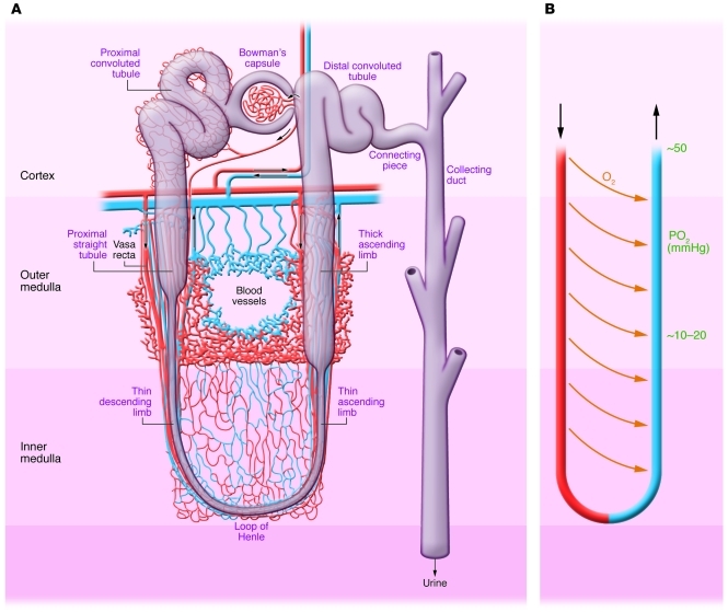
Figure 2. Normal nephron, corticomedullary oxygen gradient, and outer medullary microvascular anatomy.
(A) Anatomy of nephron with regions identified. Outer medulla vasculature is shown with capillaries in red and venous system in blue. (B) The vasa recta with countercurrent exchange of oxygen resulting in a gradient of decreasing oxygen tension.

