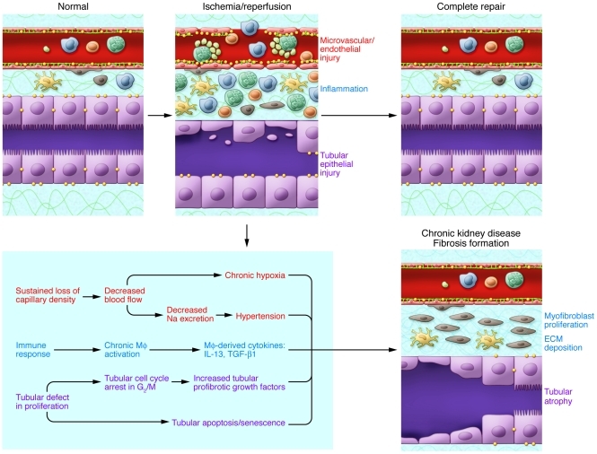
Figure 7. Abnormal repair in ischemic AKI.
Repair after AKI can result in incomplete repair and fibrotic lesions, which may result in progressive renal dysfunction. Factors including long-term hypoxia and hypertension result from chronic loss of peritubular microvessels. Sustained production of profibrotic cytokines such as IL-13, arginase, and TGF-β1 from the chronically activated macrophages (MΦ) contribute to postischemic fibrosis. Renal tubular epithelial cells also play a critical role in the development of fibrosis through fundamental changes in their proliferation processes, including cell cycle arrest in the G2/M phase. This results in a secretory phenotype that facilitates the production by the epithelial cells of profibrotic growth factors (including TGF-β1 and CTGF). Fibrogenesis is stimulated, and progression to chronic renal failure is accelerated.

