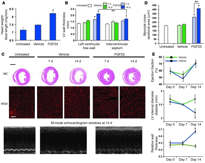
Figure 4. Intramyocardial injection of FGF23 induces LVH in mice.
(A) Intramyocardial injection of FGF23 induces a significantly increased ratio of heart weight to tibial length by day 14 (*P < 0.01, compared with vehicle). (B) Intramyocardial injection of FGF23 induces significantly increased thickness of the left ventricular free wall that is detectable at day 7 and progresses by day 14. Hypertrophy is significantly more pronounced at the injection site in the free wall compared with the interventricular septum at 14 days (*P < 0.01, compared with vehicle at corresponding date and site; **P < 0.01, comparing free wall to septum). (C) Representative gross pathology section from the cardiac mid-chamber (MC; hematoxylin and eosin stain; original magnification, ×5; scale bar: 200 μm) and WGA-stained sections from the mid-chamber free wall (original magnification, ×63; scale bar: 50 μm) demonstrate FGF23-indcued LVH at 14 days, confirmed by M-mode echocardiography. (D) Intramyocardial injection of FGF23 induces significantly increased cross-sectional surface area of individual cardiomyocytes (*P < 0.01, compared with vehicle; **P < 0.01, comparing day 14 versus 7). (E) Echocardiography at baseline and at 1 and 2 weeks after injection of FGF23 or vehicle reveals no change in ejection fraction but significantly decreased left ventricular internal diameter in diastole and increased relative wall thickness by day 14 in the mice injected with FGF23, consistent with concentric LVH (*P < 0.01, compared with vehicle). All values are mean ± SEM; n = 3 mice per group for morphological analyses; n = 100 cells per group for WGA analysis.

