Figure 3.
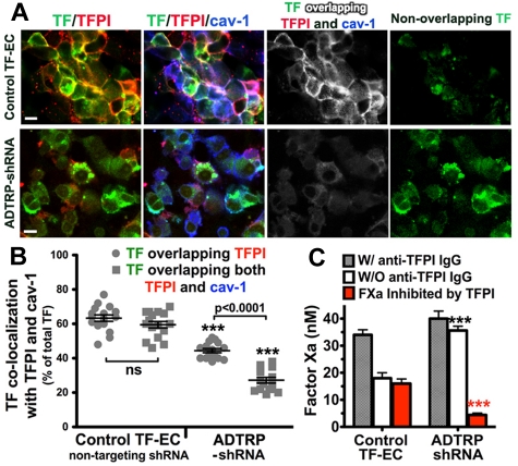
Silencing of ADTRP decreases cell surface TFPI expression and activity. (A) Immunofluorescence and confocal microscopy showing the distribution and colocalization of cell surface TFPI (Cy3, red) with YFP-TF (YFP, green) and Cav-1 (Cy5, blue; after permeabilization) on controls (nontargeting shRNA) and ADTRP shRNA-expressing TF-EC. Triple overlapping (white) and nonoverlapping TF (green) channels were generated by the Adobe Photoshop image analysis. Bars: 10 μm. (B) Scatter-plot and statistical analysis (1-way ANOVA) of the colocalization percentage between TF and TFPI, and TF/TFPI/Cav-1, out of the total MFI of TF. ***P < .001 for both sets of data in the ADTRP-shRNA group compared with control (nontargeting shRNA) TF-EC group. ns, P > .05 for the difference between the sets of data within the control experimental group. Data are mean ± SEM. (C) The inhibitory capability of cell surface TFPI against TF-FVIIa–dependent FXa generation measured in TF-EC in the presence/absence of inhibitory anti-TFPI IgG. Values are normalized to 106 cells. ***P < .001 versus control (unpaired t test). Data are mean ± SD of triplicates.

