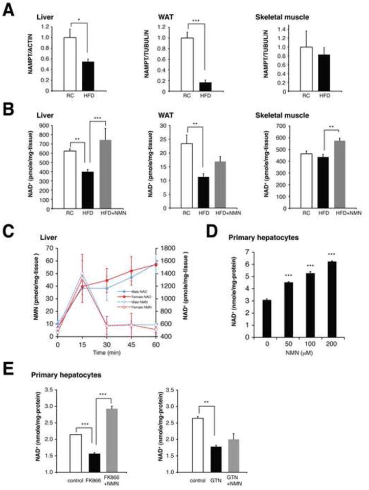
Figure 1. NMN ameliorates defects in NAMPT-mediated NAD+ biosynthesis in HFD-induced diabetic mice.

(A) NAMPT protein levels in the liver, WAT, and skeletal muscle. Female mice were fed a RC or a HFD for 6–8 months. NAMPT levels were normalized to ACTIN (liver) or TUBULIN (WAT and skeletal muscle) (n=4 to 5 mice per group). (B) Tissue NAD+ levels in the liver, WAT, and skeletal muscle from RC, HFD, and NMN-treated HFD mice (n=5 to 13 mice per group). NMN (500mg/kg body weight/day) was given intraperitoneally to HFD-fed female mice for 7 consecutive days. (C) Changes in NMN and NAD+ levels in the liver after administering a single dose of NMN to B6 mice (n=3 to 5 mice for each time point). (D and E) Intracellular NAD+ levels in mouse primary hepatocytes. Cells were treated with NMN at the indicated concentrations (D), or with enzyme inhibitors [500 nM FK866 or 100 μM gallotannin (GTN)] in the presence or absence of 100 μM NMN (E), for 4 hrs (n=3 per group). Data were analyzed by Student’s unpaired t test (A) and one-way ANOVA with the Fisher’s PLSD post-hoc test (B, D, E). All values are presented as mean ± SEM. *P < 0.05; **P <0.01; ***P < 0.001.
