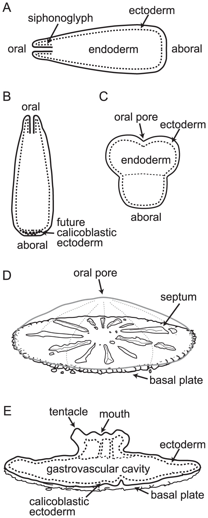
Figure 3. Schematic diagram labeling the morphological features discussed in the text and figure captions.

(A) A planula larva swimming horizontally pre-settlement has a lipid filled endoderm surrounded by a ciliated ectoderm. The siphonoglyph is an infolding of the body wall at the oral pore. Swimming direction is aboral end first. (B) When ready to settle, the planula begins searching the bottom until it encounters appropriate chemical cues. The majority of planulae then round up and settle. However, morphology is variable and specimens such as that shown in (C) are often found; these may represent larvae which have started to settle and then rejoined the plankton. (D) The planula settles on its aboral end and calcification starts in the space between the calicoblastic ectoderm and the substratum. Once a calcified plate has been laid down vertical skeletal elements (septa) start to be formed, dividing the plate into segments. (E) Six tentacles develop around the mouth of the first polyp as its column elongates.
