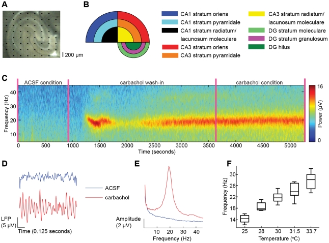
Figure 1. Local field potentials were recorded in hippocampal slices with multi-electrode arrays during ACSF and during application of carbachol.
(A) The 8-by-8 multi-electrode array covering a slice of the mouse hippocampus. Black dots are electrodes and the spacing is 200 µm. For every slice, a photograph was taken to classify the electrode location into one of the nine hippocampal subregions shown in (B). (C) Time-frequency representation of a signal in CA3 stratum pyramidale for the complete experimental recording. (D) Examples of broadband (1–45 Hz) local field potential (LFP) traces in the ACSF (blue) and carbachol (red) condition. (E) Amplitude spectra of representative signals in each condition. (F) Box-plot summaries of peak frequencies of carbachol-induces oscillations, measured at different temperatures. The frequency approaches the gamma-frequency range (>30 Hz) at physiological temperature.

