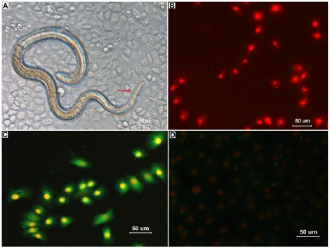
Figure 4. Observations on invasion of IECs by Trichinella spiralis infective larvae.
(A) When T. spiralis infective larvae were inoculated onto monolayers of mouse IECs in vitro, the larvae invaded the cells, and their head and body resided in the cytoplasm of the syncytia composed of numerous IECs (showed as arrow). (B) After the removal of agarose, the cell monolayer was stained with propidium iodide (PI) and observed under a fluorescence microscope. Nuclei of the damaged cells were stained intensely and uniformly red, showing the serpentine trail left by the parasite. In contrast, nuclei of the live cells not invaded by the larvae were not stained. (C) After being stained with PI, the cell monolayer was fixed and reacted with rabbit immune sera against T. spiralis ES antigens and FITC-conjugated secondary antibody as described in Materials and Methods. Green fluorescence was found in the cytoplasm of the damaged cells. (D) No fluorescence was found in the cells which were not invaded by the non-activated larvae.

