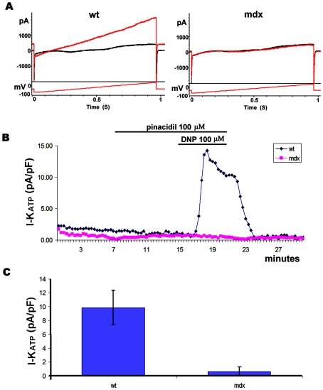
Figure 4. Current recorded from adult cardiomyocytes.
A) Wt and mdx traces primed with 100 µM Pinacidil, before and after application of DNP 100 µM in voltage clamp whole cell configuration. The voltage protocol used to elicit the current is also indicated. The red trace shows the current after 5 minutes of DNP application. B) Time course of a recording from wt and mdx cells in relation to the timing of Pinacidil and DNP application. C) KATP current quantified from the recording of 9 wt (from 3 mice) and 12 mdx (from 4 mice) cells. The current was normalized to the cell size.

