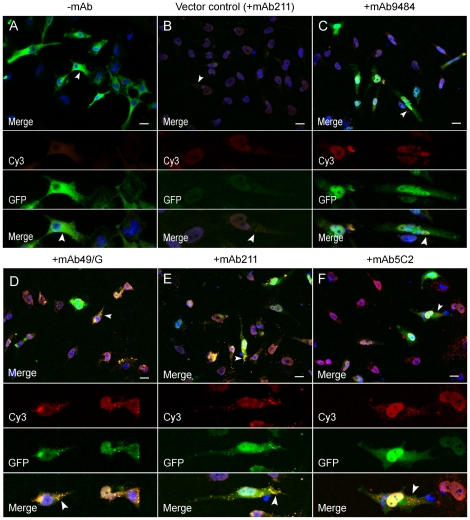
Figure 2. Immunocytochemistry with anti-mouse secondary antibodies (Cy3) displays internalization of the α-synuclein monoclonal antibodies (+mAb).
Forty-eight hours after transfection with the two α-synuclein hemi:GFP constructs, cells displayed GFP fluorescence in the whole cell soma (green) (Fig. A). Cells transfected with the constructs and treated with the α-synuclein antibodies mAb49/G and mAb211 displayed less diffuse GFP fluorescence but more localized GFP-punctae (Fig. D, E, arrows). These signals occasionally co-occurred with signals from an anti-mouse secondary antibody, indicating internalization (yellow) of the treatment antibodies (Fig. D, E, arrows). Cells treated with the mAb5C2 antibody only displayed diffuse GFP-fluorescence in the whole cell soma (Fig. F). After staining with an anti-mouse secondary antibody red punctae could be detected in the cells indicating no co-occurrence with GFP (Fig. F, arrows). The blue signal represents DAPI nuclear staining (40x magnification. Scale bar 20 µm).

