Figure 1.
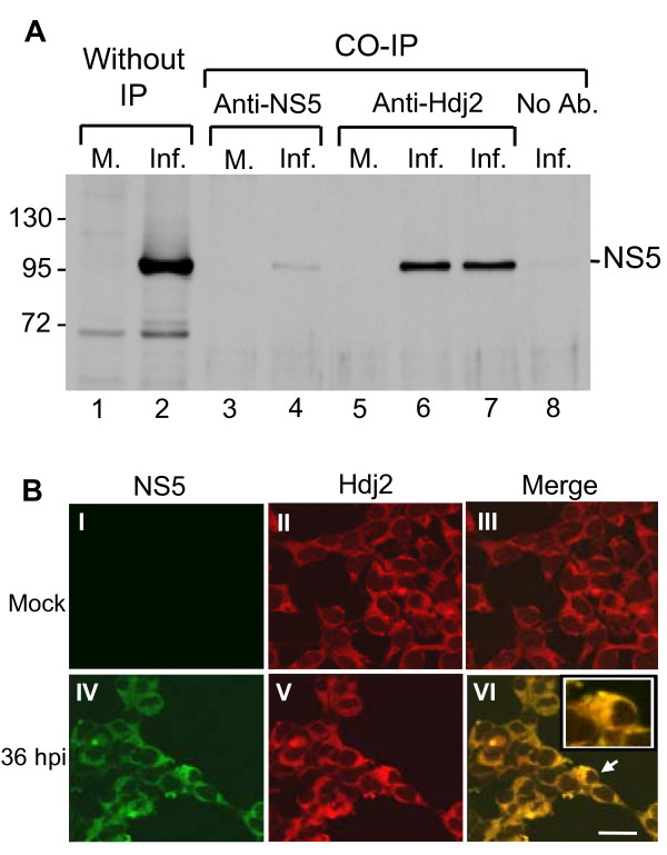
JEV viral NS5 interacts with Hdj2 in vivo. A. HEK293 cells were mock-infected or infected with JEV and harvested at 48 h post-infection. Cell lysates were subjected to co-immunoprecipitation (IP) with antibodies against NS5 or anti-Hdj2, followed by Western blot analysis for the detection of NS5. Lanes 1 and 2, Mock- and JEV-infected cell lysates; lanes 3 and 4, Co-IP with anti-NS5 antibody; lanes 5-7, Co-IP with anti-Hdj2 antibody (0.5 μg and 1 μg of anti-Hdj2 antibody was used for lanes 6 and 7, respectively); lane 8, Co-IP with no added antibody. B. Mock- or JEV-infected HEK293 cells were stained by indirect immunofluorescence using rabbit anti-NS5 polyclonal antibodies (panel I, IV), and mouse anti-Hdj2 antibody (panel II, V) and detected by FITC-conjugated goat anti-rabbit and CY3-donkey anti-mouse antibody, respectively. Panels III and VI are merged images from two previous panels. The enlarged image of the selected area marked by an arrow is given in the upper right corner of panel VI. Cells were viewed at 1000 × magnification; a 20 μm scale bar is shown.

