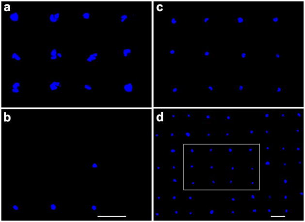
Figure 4.
Fluorescent images of HUVEC cells cultured on gold-patterned silicon oxide substrates with gold squares coated with fibronectin (a: 10× objectives), physically adsorbed REDVY (b: 10 × objectives), and covalently bound KREDVY (c: 10× objective, d: 5× objective). Image (c) was captured from image (d) in the area surrounded by the white rectangle. Scale bars are 60 μm in all images.

