
Figure 5.
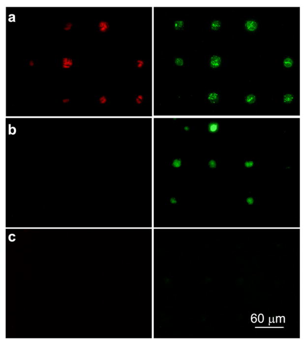
Fluorescence images of HUVEC cells on the gold patterns immobilized with (a) Fibronectin, (b) REDVY peptide, and (c) KREDVY peptide after 7 days of cell adhesion. Apoptotic cells fluoresce green, necrotic cells fluoresce red, and live cells show little or no fluorescence. Images were taken from triplicate substrates for each type of surfaces.