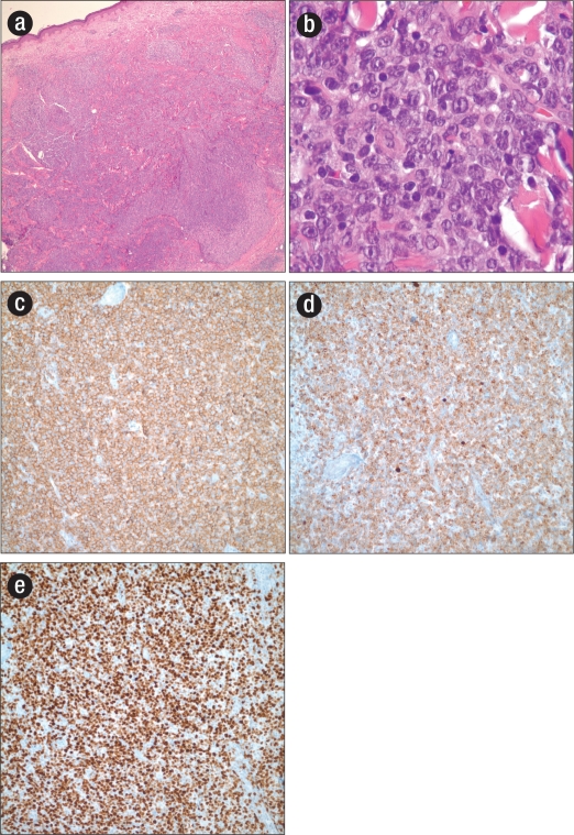
Figure 1.
(a) Hematoxylin and eosin stain showing nodular infiltration of the superficial and deep dermis by primary cutaneous diffuse large B-cell lymphoma, leg type (low power). (b) Hematoxylin and eosin stain showing large monomorphic cells with a vesicular chromatin pattern, prominent nucleoli, and frequent mitoses (high power). (c) Immunohistochemical staining for CD20. (d) Immunohistochemical staining for Bcl-2. (e) Immunohistochemical staining for MIB-1.

