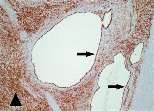
Figure 6.
Photomicrograph of the spleen (original magnification ×100) shows a representative cystic lesion with CD31 immunohistochemical stain, a vascular marker. The thin cellular lining (arrows) is strongly positive, consistent with vascular endothelium. The sinusoidal endothelial cells of the red pulp (arrowhead) also show strong positivity, as expected.

