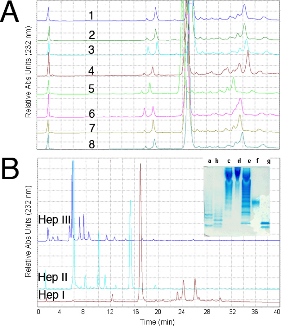
Figure 4.
Quantitative disaccharide mapping of heparins using SAX-HPLC. A. SAX-HPLC analysis of heparins #1–3 and 5–8 and LMWH #4 digested by Hep I; B. HPLC analysis of heparin #1 digested by HepI, HepII and HepIII, respectively Insert: PAGE analysis of digested heparin. Lanes a, b and c, heparin #1 digested by Hep I, II and III, respectively, d, heparin #1, e, oligosaccharide ladder (see Figure 1 legend), f, oligosaccharide standard dp10 (see Figure 1 legend), g, oligosaccharide standard dp4 (most intense band corresponds to ΔUA2SGlcNS6S-IdoA2SGlcNS6S.

