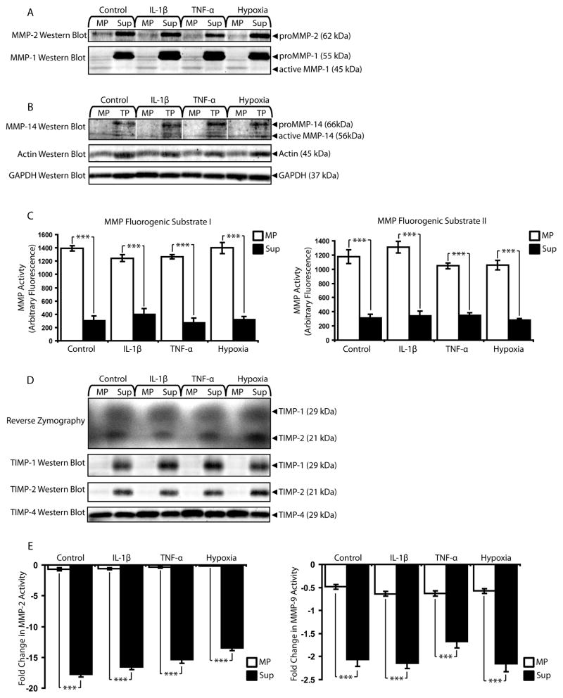
Figure 7.
MPs remain functional units of MMP activity in pathological conditions simulated in vitro with cytokines and hypoxia. (A) MMP-2 and MMP-1 western blot of MP and Sup fractions collected from microECs cultured under simulated pathological conditions (exposure to IL-1β, TNF-α, or hypoxia). (B) MMP-14 western blot of microEC MP and TP samples from control and pathological conditions. (C) MMP activity assays with MMP substrates I and II. ***p < 0.001 (D) Reverse zymography and TIMP-1, 2, and 4 western blots of MP and Sup produced in the presence of IL-1β, TNF-α, or hypoxia. (E) MMP-2 and MMP-9 inhibitor assays. ***p < 0.001 Results are means ± SD.

